Usted está aquí
Peruvian Journal of Neurosurgery
Hydrocephalus in pediatric population. Experience in the Neurosurgery Department of the Baca Ortiz Pediatric Hospital, Quito-Ecuador, 2016-2019
CARLOS MORALES T., ALICIA TORRES M., JESUS CASTRO V., JOSE BERNAL C., ALEJANDRO CASTRO S.
Abstract (Spanish) ||
Full Text ||
PDF (Spanish) ||
PDF (English)
ABSTRACT
|
Introduction: Hydrocephalus is one of the most common pathologies in the daily care of a Pediatric Neurosurgery Service, so it is important to document the context of its presentation to improve therapeutic behavior. The objective of the present study was to describe the demographic variables, as well as the type, treatment, morbidity, and mortality of patients with hydrocephalus.
Methods: Cross-sectional, retrospective, and descriptive study of children with hydrocephalus who underwent surgery in the Neurosurgery Service of the Baca Ortiz Pediatric Hospital, from January 2016 to December 2018, using the records of the clinical records from the Department of Statistics and archives of the Neurosurgery Service of our Institution.
Results: From January 2016 to December 2018, 287 patients with a diagnosis of hydrocephalus underwent surgery, presenting 63.7% as a congenital cause and 36.2% acquired; with a predominance of males (57.2%). The most common sign for which patients attended the consultation was macrocephaly (52%). Infectious dysfunctions were 10.1% vs. mechanical dysfunctions 4%. Mortality was not related to hydrocephalus, 1% of deaths were associated with systemic infection, and 0.6% with a respiratory infection.
Conclusions: Hydrocephalus in pediatric patients constitutes a frequent pathology that can be accompanied by various comorbidities, hence the importance of its timely diagnosis and adequate treatment.
Keywords: Hydrocephalus, Child, Neurosurgical Procedures, Hospitals, Pediatric. (Source: MeSH NLM)
|
Vertebral hydatidosis: case report
JOHN VARGAS U, OSMAR ORDINOLA C, EDUARDO LAOS P, ALFONSO BASURCO C.
Abstract (Spanish) ||
Full Text ||
PDF (Spanish) ||
PDF (English)
ABSTRACT
Introduction: Vertebral hydatid cyst is a rare disease with an incidence rate of 0.2-1%. In the spinal cord, it can cause pain and severe disability due to compression and instability. Magnetic resonance imaging (MRI) shows characteristic lesions in T1 and T2. The combination of medical anthelmintic treatment and surgery for cyst excision is the most used strategy.
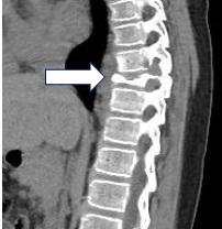
Clinical Case: A 47-year-old woman with a history of surgery for hepatic hydatidosis associated with intraoperative rupture, with a 7-month history of the disease characterized by oppressive back pain, belt-like back pain, and of increasing intensity up to 10/10, associated with paresthesia in the lower limbs and urinary retention. The MRI revealed a multicystic process in the D5 vertebral body with the invasion of the posterior mediastinum and the spinal canal. The diagnosis was confirmed serologically with IgM and IgG. A D5 corpectomy and excision of the cystic lesion were performed using a D5-D6 costotransversectomy; in addition, a D3-D4 and D6-D7 transpedicular fixation and a D5 body replacement with cylindrical mesh were performed. The evolution was favorable without evidence of recurrence one month after surgery.
Conclusion: Vertebral hydatidosis is a rare pathology that requires surgical management combined with prolonged medical treatment with albendazole. Surgery is complex because the presence of multicystic lesions makes extensive surgical resection without rupture of the cyst membrane difficult. Close monitoring of these patients allows detection of recurrence and favors its early management.
Keywords: Echinococcosis, Spinal Cord, Back Pain, Albendazole, Surgical Mesh (Source: MeSH NLM)
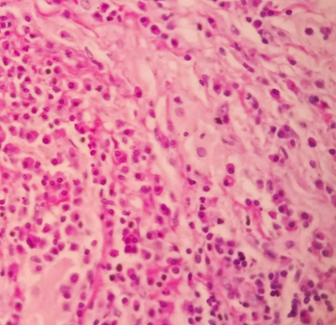
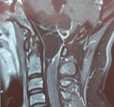
Langerhans cells histiocytosis in the dorsal spine in a pediatric patient
ANNEL MURGA V., ROBERTO BURGOS., ALFONSO BASURCO C., ELAR CARI C., JESÚS CABREJOS B., EDUARDO LAOS P., CÉSAR POLO DE LA P., JUAN SALAS G.
Abstract (Spanish) ||
Full Text ||
PDF (Spanish) ||
PDF (English)
ABSTRACT
Introduction: Langerhans cell histiocytosis (LCH) is a rare histiocytic disorder and its incidence is not exactly known. It occurs in all age groups but is more common in the pediatric population. It is characterized by single or multiple osteolytic-type lesions caused by clonal proliferation of cells histologically like Langerhans cells; its clinical presentation is heterogeneous.
Clinical case: An 11-year-old man with a 6-month history of back pain and walking limitation. Magnetic Resonance Imaging (MRI) showed a lesion of the dorsal spine in D8, D9, and D10 and flat vertebra D9 that caused spinal compression. The diagnosis was made based on the histopathological study of the vertebral body with the finding of eosinophilic granuloma, being treated with outpatient chemotherapy, external fixation with a plaster corset, and physical therapy. The clinical evolution was favorable, achieving improvement in muscle strength and walking with support at discharge.
Conclusion: Langerhans cell histiocytosis with vertebral involvement is a highly relevant pathology, despite being rare. Timely diagnosis and adequate treatment are essential since it allows to prevent or limit the spinal cord involvement caused by this pathology.
Keywords: Histiocytosis, Langerhans-Cell, Spine, Back Pain, Eosinophilic Granuloma (Source: MeSH NLM)
Successful management of ruptured cavernous malformation of the pons
JOHN VARGAS U., FERNANDO PALACIOS S., EDUARDO ROMERO V.
Abstract (Spanish) ||
Full Text ||
PDF (Spanish) ||
PDF (English)
ABSTRACT
Introduction: Brain stem cavernomas constitute 18-35% of intracranial cavernomas and have the highest bleeding rate of all brain cavernomas. Its annual rebleeding rate is 21 to 60%. Asymptomatic injuries should be treated conservatively, while symptomatic and accessible injuries surgery is recommended. Surgical resection prevents neurological deterioration caused by recurrent bleeding.
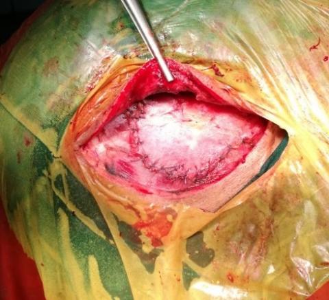
Clinical Case: We present the case of a 10-year-old female patient with headache, ataxic gait, left LV PC paresis, and vertigo. A magnetic resonance imaging of the brain (MRI) was carried out, where a ragged expansive process was evidenced in T2 in the posterior protuberance region with intra-tumoral and extra-tumoral hemosiderin deposits. Brain angiography was negative. A medial suboccipital craniotomy with a telovelar approach was performed, achieving total cavernoma resection. The patient was extubated on the 2nd postoperative day. Non-contrast brain tomography (CT) showed no acute complications, only minimal bleeding from the operative bed. Post-surgical brain MRI showed the absence of the lesion. At 7 months, she continued without motor deficit, with adequate gait and Glasgow 15 points.
Conclusion: Brain stem cavernomas are a rare disease with high morbidity. Proper patient choice, surgical approach, and time to surgery decrease post-surgical morbidity.
Keywords: Hemangioma, Cavernous, Central Nervous System, Brain Stem, Craniotomy (Source: MeSH NLM)
Experience in the surgical treatment of pituitary adenomas at the Guillermo Almenara Hospital in 2019-2020
JOHN VARGAS U, GIAN FRANCO REYES N, FERNANDO PALACIOS S, MARCO MEJIA T, JERSON FLORES C, CAMILO CONTRERAS C, MANUEL LAZÓN A, KENNET LOPEZ G, JOHN MALCA B, JOSE-DANIEL FLORES S, EDUARDO ROMERO V.
Abstract (Spanish) ||
Full Text ||
PDF (Spanish) ||
PDF (English)
ABSTRACT
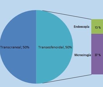
Introduction: Pituitary adenomas constitute 25% of the primary benign neoplasms of the brain and can be functional or non-functional, or depending on their size they can be microadenomas, macroadenomas, and giant adenomas. They are mainly treated by surgery using a transcranial or transsphenoidal approach.Objective: To know the experience in the surgical treatment of pituitary adenomas in the Guillermo Almenara National Hospital from January 2019 to May 2020.Methods: Descriptive, retrospective, cross-sectional epidemiological study. 84 cases of patients operated on for pituitary adenoma were found. The data was collected from the medical history and images in the hospital's PACS system. Chi-square was applied as a statistical test.Results: Of the total of patients, 50% were operated by transcranial surgery and 50% by transsphenoidal surgery. Hypertension, Cushing's disease, and acromegaly were statistically significant in favor of transsphenoidal resection. 69.05% were macroadenomas in transsphenoidal resection (TSR) and 61.90% in transcranial resection (TCR). In the TSR there were 4.76% of intraoperative complications, and in the TCR it was 19.05%. The total resection grade was greater than 50% in both groups.Conclusions: Pituitary adenomas are a frequent pathology and can be treated by transcranial or transsphenoidal approach, with good resection rates. Prospective studies are required to determine the causal relationship between the variables. Keywords: Pituitary Neoplasms, Acromegaly, Craniotomy, Endoscopy (Source: MeSH NLM)
|
Malfunction of the valvular shunting system in children. Experience in the Neurosurgery Department of the Baca Ortiz Pediatric Hospital, Quito-Ecuador, 2016-2019
JUAN ALEMÁN-IÑIGUEZ, ALICIA TORRES M, JESUS CASTRO V, JOSE BERNAL C.
Abstract (Spanish) ||
Full Text ||
PDF (Spanish) ||
PDF (English)
ABSTRACT
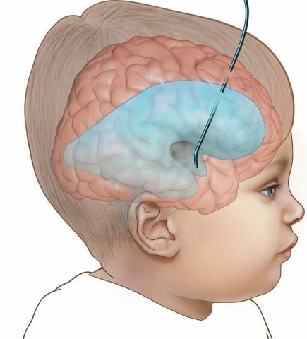
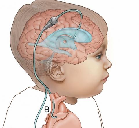
Introduction: The prevalence of valve dysfunction (VD) in pediatric centers is high. A descriptive observational study was carried out, the objective of which was to find factors: epidemiological characteristics of hydrocephalus and other derivatives of the ventricle-peritoneal shunt system (VPSS) associated with VD.Methods: All the diagnoses of VD for 3 years in the Neurosurgery Service of the Baca Ortiz Pediatric Hospital (BOPH) were collected, clinical-epidemiological variables associated with hydrocephalus and shunting were selected. VD was defined as the revision of the referral in patients using VPSS for malfunction. Multi-categorical variables and the prevalence of qualitative variables were analyzed using statistical analysis.Results: A total of 376 children were initially subjected to VPSS placement between August 2016 and August 2019. 71 patients with VD were treated, of whom 60 were included in the study; of these 48 were treated at BOPH. Infectious dysfunction was found to be more frequent in children < 1-year, mechanical dysfunction in children > 5 years (64% and 38% respectively p <0.002). Also, the permanence of the VPSS <1 year was more frequent in infectious dysfunctions and the permanence of 1 to 5 years was more related to mechanical dysfunction (72 and 46% respectively p 0.03). The distal catheter dysfunction was more important in mechanical and mixed etiology (65.41 respectively p <0.001) and that of the ventricular catheter in infectious etiology (81% p <0.001). No independent association of VD was found with the etiology of hydrocephalus, the ventricular catheter site or with the Lansky scale.Conclusions: Valvular dysfunction in pediatric hydrocephalic patients is an important complication that warrants further investigation.Keywords: Hydrocephalus, Catheters, Neurosurgical Procedures, Hospitals, Pediatric. (Source: MeSH NLM) |
Embolization of ruptured giant aneurysm of the medium cerebral artery followed by surgical evacuation of intracerebral hematoma
JOSÉ LUIS URQUIZO R, RODOLFO RODRÍGUEZ V, WALTER DURAND C, RICARDO VALLEJOS T, DANTE VALER G, JESÚS FLORES Q, GIANCARLO SAAL Z.
Abstract (Spanish) ||
Full Text ||
PDF (Spanish) ||
PDF (English)
ABSTRACT
Introduction: Early endovascular therapy of a ruptured giant aneurysm associated with intracerebral hematoma avoids the need for clipping of the aneurysm, thereby avoiding the need for greater brain retraction, edema, transient clipping, as well as the possibility of intraoperative rupture, thus achieving timely and adequate management of this pathology.
Clinical case: We present the case of a 48-year-old woman with subarachnoid hemorrhage (SAH) and intracerebral hematoma (ICH) due to dysplastic giant aneurysm of the right middle cerebral artery bifurcation (MCA) who was treated by coils embolization followed by microsurgical evacuation of the hematoma achieving a good result and a favorable clinical outcome.
Conclusion: The combined treatment by embolization with coils of a giant ruptured aneurysm of the MCA followed by the microsurgical evacuation of the ICH during the acute phase is an effective and safe alternative to single surgical management.
Keywords: Aneurysm Ruptured, Subarachnoid Hemorrhage, Hematoma, Embolization Therapeutic. (Source: MeSH NLM)
Successful endovascular management of a ruptured cerebral aneurysm in an infant patient
JOHN VARGAS U, JESÚS FLORES Q, RODOLFO RODRÍGUEZ V, WALTER DURAND C, DANTE VALER G.
Abstract (Spanish) ||
Full Text ||
PDF (Spanish) ||
PDF (English)
ABSTRACT
Introduction: Cerebral aneurysms in pediatric age are rare. In early childhood, they appear before the age of 2 years and are related to a high incidence of injuries along the middle cerebral artery, in its distal part, and in the vertebrobasilar system. The etiology can be idiopathic, traumatic, and fungal. Aneurysm obliteration should be as early as possible in patients with low surgical risk.
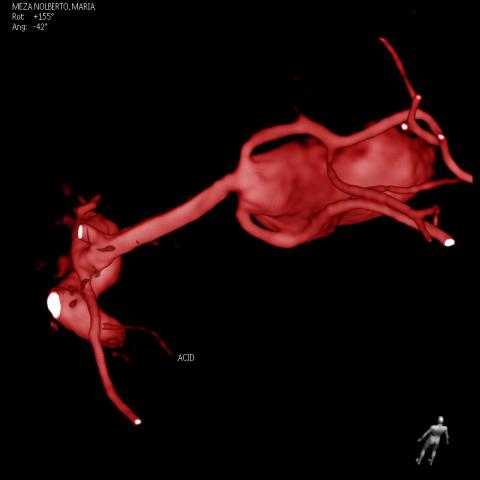
Clinical Case: The case of a 5-month-old patient with no significant medical history is presented, with a 2-day illness time, signs of irritability, vomiting, and tension in the fontanelle. A cerebral tomography was performed showing a predominantly right subcortical frontal fine subarachnoid hemorrhage and an angio-tomography (Angio-TEM) that showed an aneurysm of the anterior cerebral artery. The cerebral angiography study revealed a dissecting aneurysm of the left A2-A3 segment that involved the division of the left anterior cerebral artery into a pericallosal and marginal callus artery. Embolization was performed using 4 coils and Histoacryl® to close the parental artery. He had a seizure crisis from a left marginal callus infarction that was medically controlled. The clinical evolution was good, being discharged on the 7th day of hospitalization.
Conclusion: Pediatric cerebral aneurysms are a rare pathology and in patients with low surgical risk, such as our patient, they should be treated as soon as possible to decrease morbidity and mortality.
Keywords: Intracranial Aneurysm, Cerebral Angiography, Infant, Embolization Therapeutic. (Source: MeSH NLM)

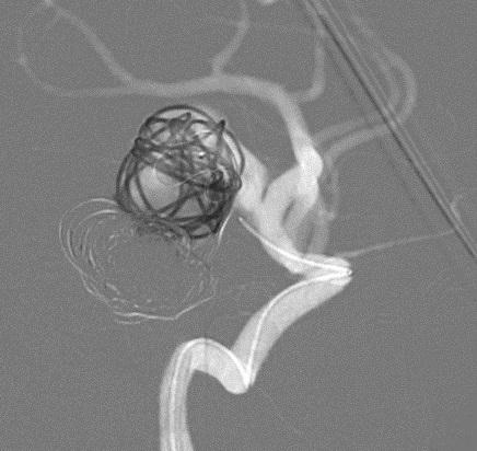
Endovascular treatment of a large, embolized, unruptured cerebral aneurysm that presented rechanneling
YHAKELIN ESPINOZA C, JOHN VARGAS U, RODOLFO RODRÍGUEZ V, WALTER DURAND C, JESÚS FLORES Q, DANTE VALER G, GIANCARLO SAAL Z.
Abstract (Spanish) ||
Full Text ||
PDF (Spanish) ||
PDF (English)
ABSTRACT
Introduction: Endovascular treatment of ruptured and non-ruptured brain aneurysms is an appropriate option in most patients. In narrow neck aneurysms, treatment with coils is sufficient, but in those with wide necks, the use of stent-assisted coils or flow diverters is necessary. Endovascular treatment of intracranial aneurysms is effective and provides complete occlusion in up to 85% of cases, however, there are cases of incomplete obliteration or recanalization that require treatment.Clinical Case: A 79-year-old woman with a history of high blood pressure and embolization with coils of an unruptured cerebral aneurysm of the left posterior communicating artery, 5 years ago, Raymond Roy I in the immediate postoperative period. She has been presenting with right hemiparesis and III left cranial nerve palsy for 1 year. An angiotomography (angioCT) showed recanalization of the aneurysm, so it was decided to perform an embolization with coils and stents, achieving complete occlusion of the aneurysm (Raymond Roy I). In the following weeks, the patient regained strength in the right hemibody and recovered function of the III left cranial nerve.Conclusion: Recanalization of aneurysms undergoing endovascular treatment is infrequent, less than 10%, and is associated with complex aneurysms. In these cases, the use of advanced or combined endovascular techniques is recommended to achieve total closure of the aneurysm and decrease the associated morbidity and mortality rate.Keywords: Intracranial Aneurysm, Endovascular Procedures, Stent, Embolization Therapeutic. (Source: MeSH NLM) |
Experience in diagnostic and treatment of central nervous system tumors in children less than 2 years at the Baca Ortiz Pediatric Hospital, Quito-Ecuador, 2016-2019
CARLOS FLORES E, ALICIA TORRES M, JOSE BERNAL C, JESUS CASTRO V.
Abstract (Spanish) ||
Full Text ||
PDF (Spanish) ||
PDF (English)
ABSTRACT
|
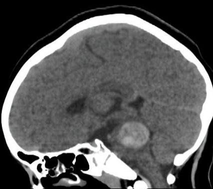
Introduction: Tumors of the central nervous system (CNS) in children between 0 and 2 years of age are infrequent, so their diagnosis and treatment constitute an important challenge for every pediatric neurosurgical center, to which is added the comorbidities typical of this age that condition the responsibility of improving therapeutics to obtain better survival. The objective of this study was to describe the diagnostic and therapeutic experience of a neurosurgical Department of national reference, in relation to neoplasms of the central nervous system in children under two years of age, as well as to establish comorbidity and prognosis.
Methods: Cross-sectional, retrospective and descriptive study that includes all patients under 2 years of age who were diagnosed with a neoplastic lesion of the central nervous system, attended from January 01, 2016 to July 01, 2019 at the Baca Ortiz Pediatric Hospital from the city of Quito in Ecuador.
Results: CNS tumors in children between 0 and 2 years old corresponded to 5.09%, with irritability being the most frequent reason for consultation with 62.5%. Also, 75% of the neoplasms were located at supratentorial level with a 1: 1 intra / extra-axial ratio. Neuroblastoma and choroid plexus tumors were the most frequent histopathological diagnoses. Mortality had a percentage of 50%.
Conclusions: CNS tumors in children between 0 and 2 years are not frequent, the location is predominantly supratentorial and the prognosis for life depends on the histopathological type. Radiation therapy is an option, although surgery for resection is the basis of treatment.
Keywords: Central Nervous System Neoplasms, Comorbidity, Prognosis. (Source: MeSH NLM)
|

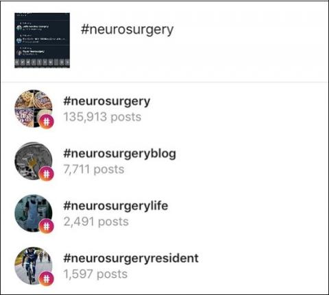
Use of social media networks in Neurosurgery
EMIL ZHALMUKHAMEDOV
Abstract (Spanish) ||
Full Text ||
PDF (Spanish) ||
PDF (English)
ABSTRACT
|
Neurosurgery as a medical discipline has always been on the cutting edge of technology and the latest advancements. It’s not a surprise why modern neurosurgeons actively utilize various social media platforms, in order to share the latest news or showcase interesting cases they face daily. From Twitter to LinkedIn neurosurgical professionals of the world share the knowledge and connect with each other in order to advance their skills to the next level. A thought-provoking content is constantly shared and spark many interesting conversations and contribute to the modern data of neurosurgical field. Keywords: Social Media, Neurosurgery, Graduate Medical Education (source: MeSH NLM)
|
Isolated intracranial Rosai-Dorfman disease: case report
JORGE ZUMAETA S, FERNANDO PALACIOS S, WILLIAM ANICAMA L, CLAUDIA BURGOS J.
Abstract (Spanish) ||
Full Text ||
PDF (Spanish) ||
PDF (English)
ABSTRACT
Introduction: Rosai-Dorfman disease is a pathology of histiocytic, proliferative, idiopathic and benign type characterized by sinus histiocytosis and massive lymphadenopathy. The most frequent clinical presentation is painless bilateral cervical lymphadenopathy. Extra-nodal involvement occurs in 43% of cases and central nervous system (CNS) involvement in 4%. CNS involvement is more common in men and manifests itself as a mass in the cranial dura, which may or may not be associated with lymph node involvement.
Clinical case: We present the case of a 51-year-old woman with a history of sinusitis, with a clinical picture of holo-cranial headache, associated with periods of disorientation and ideomotor apraxia. MRI showed a right parieto-occipital extra-axial lesion, contrast sensor with implantation in the cranial dura. A right parietal craniotomy was performed with subtotal resection of the lesion. The pathological anatomy was reported as Rosai-Dorfman disease of meninges. The evolution after surgery was favorable with remission of symptoms.
Conclusion: Rosai-Dorfman disease should be within the differential diagnosis of lesions based on implantation in the dura. Its diagnosis is eminently histological. Although there is no specific therapy, surgical removal is the most effective treatment. Adjuvant therapies such as steroids and radiation can help control residual or recurrent disease.
Keywords: Histiocytosis Sinus, Lymph Nodes, Dura Mater, Meninges, Craniotomy (source: MeSH NLM)
Enterogenous cyst of the posterior fossa: a case report
JOSÉ LEÓN P, FERNANDO ROMERO F, EUTEMIO MEDINA M, DIANA RIVAS F, LUIS ANTONIO T.
Abstract (Spanish) ||
Full Text ||
PDF (Spanish) ||
PDF (English)
ABSTRACT
Introduction: The intracranial enterogenous cyst is a benign cystic tumor lesion of very rare frequency worldwide. These cysts can occur at any level of neuroaxis and their malignant transformation is rare. The case of a 32-year-old woman with a cystic lesion in the posterior fossa at the level of the occipitocervical junction is presented.
Clinical case: A 32-year-old woman with a history of tumor surgery in the posterior fossa 4 years ago, with a clinical picture of chronic headache and quadriparesis. The magnetic resonance imaging of the cranio-cervical junction showed a cystic lesion at the level of the posterior fossa. A suboccipital craniectomy was performed with removal of the posterior arch of C1 and excision of the tumor. The histopathological study confirmed the diagnosis of enterogenic cyst.
Conclusion: The enterogenic cyst of the nervous system is an uncommon, benign pathology that can recur. The definitive diagnosis will be made by biopsy of the tumor piece.
Keywords: Cysts, Biopsy, Craniotomy, Central Nervous System, Headache Disorders (source: MeSH NLM)
Minimally invasive surgery using Mini-pterional Interfascial Approach for the Clipping of a Ruptured Aneurysm of the right Carotid Bifurcation at the Dos de Mayo National Hospital, Lima-Peru
JOSÉ LUIS ACHA S, HÉCTOR YAYA-LOO, DAVID YABAR B, JULIO QUISPE-DEXTRE, DIANA HERRERA-ORELLANA, JESÚS PASACHE-JUAREZ
Abstract (Spanish) ||
Full Text ||
PDF (Spanish) ||
PDF (English)
ABSTRACT
|
Introduction: Carotid bifurcation aneurysms represent 2 to 9% of intracranial aneurysms1. They can be treated by endovascular or microsurgical techniques3, the latter being the most complex1-4 and used with low frequency in this type of aneurysm7.
Clinical case: A 57-year-old woman with a ruptured aneurysm of the right carotid bifurcation, incidentally, another small unruptured aneurysm of the left posterior communicating was found. She underwent a mini-pterional interfascial approach and aneurysm clipping with satisfactory results and a favorable evolution.
Conclusion: Aneurysms of the carotid bifurcation are not frequent; this is a non-common case due to the age of 57 years. The technique performed allowed the identification of the aneurysm, as in the classical pterional approach, but trying to minimize morbidity. The patient did not present post-surgical complications despite the risk factors presented, such as unbroken contralateral aneurysm, edema due to subarachnoid hemorrhage and arterial hypertension.
Keywords: Aneurysm, Ruptured, Subarachnoid Hemorrhage, Hypertension. (Source: MeSH NLM)
|
Minipterional interfascial approach for microquirurgical treatment of ruptured and unruptured anterior circulation aneurysms. Initial experience in the Dos de Mayo National Hospital in Lima - Peru
JOSÉ L. ACHA S, HÉCTOR YAYA-LOO, DAVID YABAR B, RITA LOPEZ C
Abstract (Spanish) ||
Full Text ||
PDF (Spanish) ||
PDF (English)
ABSTRACT
|
Objective: To provide information on the experience in the management of ruptured and non-ruptured aneurysms of the anterior circulation through the minipterional interfascial approach, to describe the technique, clinical, surgical results, complications and advantages.
Methods: A retrospective observational study was conducted, from January to December 2018. Of 59 patients with ruptured and non-ruptured aneurysms, 33 were using a minipterional craniotomy. Clinical variables, location, complications and surgical results were analyzed.
Results: In total, there were 33 patients operated by a minipterional craniotomy, 35 aneurysms were clipped: 14 MCA (40%), 13 PComA (37%), 6 AComA (17%), 1 bifurcation of ICA (2%), 1 Choroid artery (2%). Of the total, 11 were men (33%), 22 women (66%). The Hunt and Hess of admission: I in 16 cases (48%), II in 11 cases (33%) and III in 6 cases (18%). There were 3 intraoperative ruptures and 8 radiological clinical vasospasms. Rankin's scale at discharge was: Rankin 0 in 2 patients (6%), Ranking 1 in 11 patients (33%), Rankin 2 in 10 patients (30%), Rankin 3 in 2 patients (6%), Rankin 4 in 1 patient (3%), Rankin 5 in 1 patient (3%) and Rankin 6 in 1 patient (3%).
Conclusions: The Minipterional craniotomy is reliable, less invasive, it maintains the advantages of the pterional approach but avoids greater exposure of the parenchyma and tissue manipulation. Aneurysms of the anterior circulation, ruptured and unruptured, can be treated safely and effectively with limited bone extraction, good cosmetic results and good temporomandibular function.
Keywords: Intracraneal Aneurysm, Craniotomy, Surgical Instruments. (source: MeSH NLM
|