Pituitary abscess: A case review
MARCO CHIPANA S, SANDY CABALLERO M, MANUEL CUEVA N, PEDRO SOTO P.
Abstract (Spanish) ||
Full Text ||
PDF (Spanish) ||
PDF (English)
ABSTRACT
|
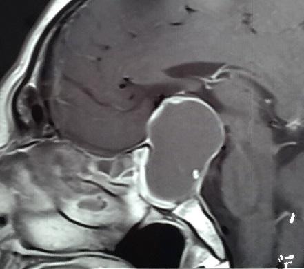
Introduction: Pituitary abscess is an intrasellar infection that generates a clinical picture like any pituitary tumor. Its origin can be of hematogenous origin or by infection of a nearby site or pre-existing lesion.
Clinical case: The case of a 38-year-old male patient with progressive headache, vomiting and visual impairment is presented. It was evaluated with magnetic resonance of the sellar region. Transsphenoidal surgery was performed and the infusion of intravenous antibiotic therapy continued.
Conclusion: Pituitary abscess is an uncommon infectious pathology whose diagnosis requires clinical and radiological evaluation. Surgery is usually necessary for diagnostic confirmation and treatment.
Keywords: Brain Abscess, Pituitary Neoplasms, Pituitary Disease (source: MeSH NLM)
|
Single-session curative embolization of a ruptured supratentorial arteriovenous malformation: Case report
ROMMEL RODRIGUEZ B, JESUS FLORES Q, WALTER DURAND C, GIANCARLO SAAL-ZAPATA, RODOLFO RODRIGUEZ V.
Abstract (Spanish) ||
Full Text ||
PDF (Spanish) ||
PDF (English)
ABSTRACT
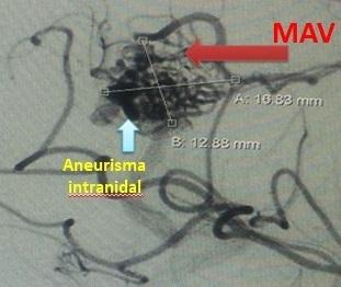
Introduction: Microsurgery has been the gold standard for the treatment of arteriovenous malformations (AVM), however, endovascular therapy has emerged as an option in recent years.
Clinical case: We present the case of a previously healthy 45-year-old woman that presented with a ruptured AVM treated successfully with curative embolization.
Conclusion: Endovascular treatment is a feasible and safe option for the treatment of arteriovenous malformations.
Keywords: Arteriovenous Malformations, Embolization, Therapeutic, Cerebral Hemorrhage. (source: MeSH NLM)
Aqueductoplasty with stent placement by neuroendoscopy for the management of a cyst of the IV ventricle in a case of neurocisticercosis
JOHN VARGAS U, JERSON FLORES C, JAIME ROJAS V, FERNANDO PALACIOS S.
Abstract (Spanish) ||
Full Text ||
PDF (Spanish) ||
PDF (English)
ABSTRACT
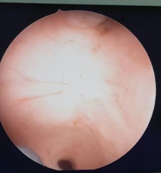
Introduction: The most common parasitic infection of the central nervous system is neurocysticercosis which in 20% of cases is intraventricular, the fourth ventricle being one of the most frequent locations. These lesions cause obstructive hydrocephalus that requires the placement of a ventricle-peritoneal shunt (DVP) whose dysfunction rate is high due to the presence of adhesions and the formation of multiple compartments being the cyst isolated from the IV ventricle one of the most difficult treatment.
Clinical Case: We present the case of a 34-year-old patient with a history of neurocysticercosis who received antiparasitic treatment and the placement of a DVP, presenting multiple dysfunction with severe dilatation of the IV ventricle. It was decided to perform an aqueductoplasty and the placement of a stent in the Silvio aqueduct followed by the placement of a new left frontal DVP, after removing the DVP with parietooccipital “Y” connector and left posterior fossa. Tomographic control at week showed a decrease in ventricular size, catheters in an adequate position, clinical improvement and good patient evolution.
Conclusion: Endoscopic aqueductoplasty with stent placement in the Silvio aqueduct is an effective measure in cases of isolated cyst in the fourth ventricle, achieving good clinical and imaging evolution compared to other therapeutic options.
Keywords: Neurocysticercosis, Hydrocephalus, Fourth Ventricle, Cerebral Aqueduct, Stents. (source: MeSH NLM)
Endovascular management of non-ruptured intracranial aneurysms with flow diverter devices. First experience in Peru.
GIANCARLO SAAL-ZAPATA, WALTER DURAND S, RICARDO VALLEJOS T, DANTE VALER G, JESÚS FLORES Q, RODOLFO RODRIGUEZ V.
Abstract (Spanish) ||
Full Text ||
PDF (Spanish) ||
PDF (English)
ABSTRACT
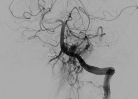
Introduction: Endovascular treatment of non-ruptured intracranial aneurysms with flow diverter devices is a technique that currently has many indications for its use.
Objectives: Determine the clinical characteristics of patients, angiographic characteristics of the aneurysms, occlusion rate at 6 months and 1 year follow-up and complications associated to the deployment of flow diverter devices in the treatment of non-ruptured intracranial aneurysms.
Methods: We present a retrospective review of consecutive cases treated with flow diverters at our institution.
Results: Since October 2012 to April 2017, twenty-one patients were treated with a total of 29 non-ruptured aneurysms. Twenty six aneurysms (90%) were located in the anterior circulation and three aneurysms were located in the posterior circulation (10%). We employed 22 flow diverters (SILK = 9, FRED = 13). Fifty percent of the aneurysms were located in the paraclinoid segment of the internal carotid artery, followed by 28% located in the cavernous segment.
Globally, fifty eight percent of the patients were cured. There were three patients with persistence of the aneurysms and five complications: three carotid thrombosis, one migration and one mal-apposition of the stent. All this complications were and remain asymptomatic. Mortality rate in this series was zero percent.
Discussion: The use of flow diverter devices is a new technique for the treatment of non-ruptured intracranial aneurysms at our institution, with adequate rates of aneurysm occlusion.
Keywords: Intracranial Aneurysm, Stents, Cerebral Angiography. (source: MeSH NLM)
Curative embolization of dural arteriovenous fistula using Onyx. Case report
GIANCARLO SAAL-ZAPATA, WALTER DURAND C., RODOLFO RODRIGUEZ V.
Abstract (Spanish) ||
Full Text ||
PDF (Spanish) ||
PDF (English)
ABSTRACT
Introduction: Dural arteriovenous fistulas (DAVF) are vascular lesions that require a high suspicious index for its diagnosis, because of their wide variety of clinical manifestations, moreover, invasive procedures such as digital subtraction angiogram (DSA) is the gold standard and is required for its diagnostic confirmation.
Clinical presentation: We report the case of a previously healthy 44-year-old young woman coming from the highlands of Peru, with the diagnosis of spontaneous intracranial hemorrhage, not treated at her local hospital. She was transferred to the capital city for complementary studies and possible treatment. DSA revealed a dural arteriovenous fistula, with feeder from the middle meningeal artery and venous cortical reflux, being catalogued as a Cognard grade IV/Borden III DAVF, we decided to perform an embolization of the fistulous communication with the non-adherent liquid embolic agent Onyx, with good clinical evolution. Immediate post embolization DSA and 6-month follow-up control DSA showed total absence of the fistula, being the patient catalogued as cured.
Conclusion: Initial diagnostic suspect, digital subtraction angiogram confirmation, an accurate study of the anatomy and the exact location of the fistulous communication are factors that favor the cure in these patients. The treatment in a specialized center that counts with the adequate infrastructure and with trained staff is vital to achieve cure.
Keywords: Fistula, Meningeal Arteries, Intracranial Hemorrhages, Angiography Digital Subtraction. (source: MeSH NLM)
Endovascular management of craniospinal junction arteriovenous fistula: Case report
JAFETH LIZANA T, GIUSEPPE ROJAS P, RODOLFO RODRIGUEZ V, WALTER DURAND C, GIANCARLO SAAL Z.
Abstract (Spanish) ||
Full Text ||
PDF (Spanish) ||
PDF (English)
ABSTRACT
Introduction: Craniocervical arteriovenous fistulas are rare entities. A high index of suspicious is required for its diagnosis. Surgery, embolization or both are available treatments.
Clinical case: Herein, we present the case of a young woman who presented with syncope. Additional studies revealed the presence of a craniocervical junction AVF. Endovascular treatment was performed in two opportunities achieving the obliteration of the fistulous communication.
Conclusion: Embolization is a safe and feasible treatment in cases of craniospinal AVFs, with good clinical and radiological results.
Keywords: Arteriovenous Fistula, Embolization, Therapeutic, Foramen Magnum. (source: MeSH NLM)
Curative transvenous embolization of a challenging ruptured thalamic arteriovenous malformation using Onyx: Case report.
PAUL CARRANZA V, WALTER DURAND C, RICARDO VALLEJOS T, DANTE VALER G, JESÚS FLORES Q, GIANCARLO SAAL-ZAPATA, RODOLFO RODRÍGUEZ V.
Abstract (Spanish) ||
Full Text ||
PDF (Spanish) ||
PDF (English)
ABSTRACT
Introduction: Arteriovenous malformations (AVM) are typically treated by arterial approach. In selected cases, venous embolization is an option with good results.
Clinical case: We present the case of a ruptured thalamic arteriovenous malformation treated successfully with transvenous embolization using Onyx, achieving complete obliteration of the nidus.
Conclusion: In selected cases, transvenous embolization is an option for the treatment of arteriovenous malformations
Keywords: Arteriovenous Malformation, Arteries, Embolization, Therapeutic, Polyvinyls. (source: MeSH NLM)
Revascularization with first generation bypass plus carotid reconstruction with tandem clipping of complex paraclinoid aneurysm. First case in the Dos de Mayo National Hospital in Lima - Peru.
JOSÉ LUIS ACHA S, HÉCTOR YAYA-LOO, PEDRO SOTO P, JORGE MURA C, JOAQUÍN CORREA, DAVID YABAR B.
Abstract (Spanish) ||
Full Text ||
PDF (Spanish) ||
PDF (English)
ABSTRACT
|
Introduction: Cerebral aneurysms are formed due to a weakness in the arterial wall, this cause a dilation and the subsequent risk of rupture, conditioning subarachnoid hemorrhage, being one of the causes of cerebrovascular disease with higher risk of morbidity or death. We presented the first brain revascularization microsurgery for the treatment of a complex cerebral aneurysm performed at the Dos de Mayo National Hospital.
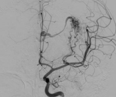
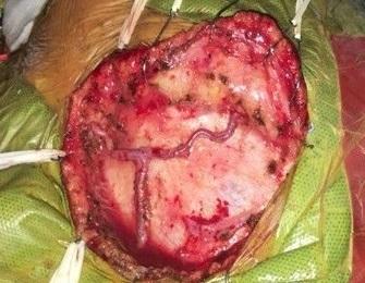
Clinical case: Woman with a ruptured left paraclinoid giant aneurysm, from Piura, who underwent revascularization by means of an extra-intracranial first generation bypass, between superficial temporal artery and silvian branch of M2, using a mini-interfascial approach, sphenoid wing drilling, anterior extradural clinoidectomy, opening of the canal, distal dural ring and tandem clipping for reconstruction of the carotid artery, excluding the complex aneurysm, was the surgical strategy chosen.
Conclusion: Post-surgical evolution was favorable, with radiological evidence of complete tandem clipping and permeability of the internal carotid and bypass.
Keywords: Intracranial Aneurysm, Cerebral Revascularization, Carotid Arteries, Microsurgery. (source: MeSH NLM
|
Mini-pterional craniotomy for clipping of anterior circulation Aneurysms
JERSON FLORES C, ALFREDO FUENTES-DAVILA M †, WESLEY ALABA G.
Abstract (Spanish) ||
Full Text ||
PDF (Spanish) ||
PDF (English)
ABSTRACT
Objectives: The pterional approach is the most common approach in vascular neurosurgery, but in recent years has been an increasing interest in minimally invasive surgery or keyhole surgery. We present preliminary results from the use of Mini-pterional Craniotomy in surgery for cerebral aneurysms performed in the Cayetano Heredia Hospital in 2009.
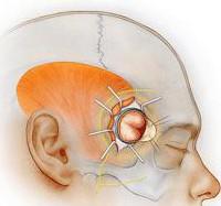
Methods: We performed a Mini-pterional Craniotomy of 2.5 x 3 cm from a curved incision of approximately 5-6 cm behind the hairline and centered on the orbitofrontal and posterior aneurysm clipping, in patients with PComA, bifurcation ICA and MCA aneurysms, during the late period, Hunt and Hess I-III, without edema, vasospasm or associated hematoma.
Results: From January to December 2009 six patients were operated by Mini-pterional Craniotomy, 4 with PComA aneurysms (66%), 1 with ICA (17%) and 1 with MCA (17%). All were operated on late and Hunt and Hess was I in 3 cases (50%) III in 2 cases (33%) and II (17%). There were no operative complications and the outcome was favorable in most cases: Rankin 1 (50%) and Rankin 2 (33%).
Conclusions: Mini-pterional Craniotomy is a minimally invasive surgery for brain aneurysms, which maintains the advantages of the standard pterional approach, but it minimizes the exposure of brain parenchyma and soft tissue manipulation. It is a valid surgical alternative in selected cases, mainly from PComA and MCA aneurysms.
Key words: Intracranial Aneurysm, Craniotomy, Minimally Invasive Surgical Procedures. (source: MeSH NLM)
Epidemiological, clinical and laboratory profile of patients submitted to micro-surgical treatment by multiple aneurysms at the Guillermo Almenara Hospital
JOHN F. VARGAS U, FERNANDO PALACIOS S, ALFREDO TUMI F, DANIEL FLORES S, JOSÉ GARCÍA V, JORGE ROMERO V, JORGE ZUMAETA S, JOSÉ URQUIZO R.
Abstract (Spanish) ||
Full Text ||
PDF (Spanish) ||
PDF (English)
ABSTRACT
|
Objective: Multiple aneurysms are common in patients with subarachnoid hemorrhage, their characteristics influence the prognosis of the patient, so their study and analysis are relevant. The objective of this study was to know the epidemiological, clinical and lab characteristics of patients with multiple aneurysms in a surgical treatment in the Guillermo Almenara Irigoyen National Hospital (HNGAI) from January 2010 to August 2017.
Methods: This study was descriptive, cross-sectional retrospective study with epidemiological type. We found 311 cases of patients with aneurysm clipping procedure of which 57 corresponded to a multiple aneurysm.
Results: Of the total number of patients, 71.93% were 50 or older, 75.44% were women and 82.46% were from the coast of the country. It was found that 28% of the patients had previous hypertension. 64.91% of patients had a Glasgow scale of 14-15 on admission, 47.37% were in the Hunt Hess II stage and 45.62% in the Fisher IV Scale. The most frequent location of the multiple aneurysms was the combination of middle cerebral artery - middle cerebral artery and the combination of middle cerebral artery - posterior contralateral communicating artery.
Conclusions: Multiple aneurysms are a frequent pathology related to female sex, advanced age and history of hypertension. The predominant affectation is the middle cerebral artery, bilaterally.
Keywords: Subarachnoid Hemorrhage, Intracranial Aneurysm, Middle Cerebral Artery. (source: MeSH NLM)
|
Severe intracranial hypertension treated with 7.5% hypertonic solution in a subarachnoid hemorrhage case by rupture of aneurysm
OSCAR SALDARRIAGA R, ELAR CARI C, GRACIELA NUÑEZ Z.
Abstract (Spanish) ||
Full Text ||
PDF (Spanish) ||
PDF (English)
ABSTRACT
Introduction: All patients with subarachnoid hemorrhage (SAH) admitted to our department are managed in intensive care unit; in the last years there has been a tendency to treat aneurysms in early stage by craniotomy or embolization, however the clinical evolution of patients is difficult in some cases because the neurosurgical critical care staff face with cerebral edema, intracranial hypertension, cerebral hypoperfusion and cerebral vasospasm among others but this early treatment has reduce the rate of rebleeding.
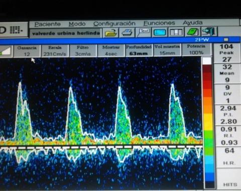
Clinical case: We report the case of a 52-year-old woman with a diagnosis of subarachnoid hemorrhage who was monitored with an intracranial pressure (ICP) sensor and transcranial Doppler (TCD) and who received treatment with hypertonic solution at 7.5% presenting a good evolution.
Conclusion: This case show us how the clinical monitoring, ICP, TCD as well as treatment with hypertonic saline solutions are good complement to take correct decisions in shortest time, because “time is brain”.
Keywords: Subarachnoid Hemorrhage, Intracranial Pressure, Hypertonic Solutions. (source: MeSH NLM)
Aneurysm associated with fenestration of Basilar artery treated by embolization with coils
ZINDYA G. BARRIENTOS M, RODOLFO RODRIGUEZ V, WALTER DURAND C, RICARDO VALLEJOS T, DANTE VALER G, JESUS FLORES Q.
Abstract (Spanish) ||
Full Text ||
PDF (Spanish) ||
PDF (English)
ABSTRACT
|
Introduction: Aneurysms of the vertebrobasilar junction are rare, but when present, they are often associated with fenestration of the basilar artery. Frequently, the endovascular treatment is the first choice due to the complex anatomy of the posterior fossa.
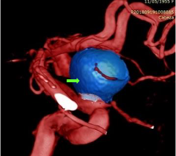
Clinical case: A 68-year-old man from Lima, who denies important antecedents, was admitted to the emergency unit with intense holocranial headache, diplopia, posterior cervical pain and vomiting, without loss of consciousness. The neurological exam showed: score of 15 in the Glasgow coma scale (GCS), no pupillary alteration, no motor or sensitivity deficit, palsy of the left sixth cranial nerve and Hunt-Hess grade II. For decision making, the patient underwent digital subtraction angiography (DSA) through the right femoral artery with 3D reconstruction (03-08-2018) which showed evidence of fenestration of the basilar artery associated with saccular aneurysm. An aneurysm coil embolization was performed without complications. The patient was discharged maintaining diplopia, with paralysis of the left sixth cranial nerve, but without any other complaints or neurological symptoms.
Conclusion: Fenestrated basilar artery aneurysms are rare and complex vascular diseases and their treatment improved with the advent of the 3D angiography and the development of the endovascular techniques. The endovascular treatment by coil embolization is the first option although other endovascular techniques have also been described.
Keywords: Intracranial Aneurysm, Basilar Artery, Endovascular Procedures. (source: MeSH NLM)
|
Penumbra Coil and Stent: An option in large aneurysms of width neck
JOHN VARGAS U, RODOLFO RODRÍGUEZ V, WALTER DURAND C, JESÚS FLORES Q, DANTE VALER G, RICARDO VALLEJOS T.
Abstract (Spanish) ||
Full Text ||
PDF (Spanish) ||
PDF (English)
ABSTRACT
Introduction: Endovascular treatment of cerebral aneurysms is an appropriate option. In patients with wide neck can be chose embolization with coils with stent assisted or flow diverter. In complex aneurysms, endovascular treatment has incomplete occlusion rates, so alternatives are sought as the Penumbra® coil, which is a thicker coil, therefore it has a potential effect of being more efficient for packaging and more cost effective, because better packaging is achieved with fewer coils and faster.
Clinical case: Presents itself the case of a 48-year-old patient, a time of illness of 8 months, with a decreased visual acuity in the left eye, sporadic headache, oppressive and global type with AVS 4/10. The cerebral angiography evidenced a saccular aneurysm of the ophthalmic segment of the left internal carotid of 15.04x11.53mm, with a 9.78mm wide neck, not broken, Barami type IA. They decide to embolize with 7 Penumbra® coils with previous placement of an LVIS® stent, achieving adequate compaction and total occlusion of the aneurysm as evidenced by immediate postoperative angiography at 6-month control, yielding the patient's clinic without any complications.
Conclusion: Penumbra® coils are an efficient alternative and cost effective of embolization in large aneurysms, and the use of stent is an ideal aid in cases of associated wide neck.
Keywords: Intracranial Aneurysm, Cerebral Angiography, Stents, Embolization Therapeutic. (source: MeSH NLM)
Endovascular treatment with coil Penumbra of a large aneurysm of the right ophthalmic segment
GONZALO ROJAS D, RODOLFO RODRÍGUEZ V, WALTER DURAND C, RICARDO VALLEJOS T, DANTE VALER G, JESÚS FLORES Q, GIANCARLO SAAL-ZAPATA
Abstract (Spanish) ||
Full Text ||
PDF (Spanish) ||
PDF (English)
ABSTRACT
Introduction: Coiling is the most common endovascular technique used for the treatment of aneurysms. Different types are available and Penumbra coils are a new option in the endovascular armamentarium.
Case report: We report the case of 63year-old female with a ruptured type IA paraclinoid aneurysm according to Barami classification, treated with 3 Penumbra coils successfully.
Conclusion: Penumbra coils seems to be an adequate option in cases of large and ruptured aneurysms of anterior circulation.
Keywords: Aneurysm Ruptured, Endovascular Procedures, Female (source: MeSH NLM)
CSF leakage and bone erosion caused by cysts in basal cistern neurocysticercosis treated by endoscopy
JOHN MALCA B, JOSE-DANIEL FLORES S.
Abstract (Spanish) ||
Full Text ||
PDF (Spanish) ||
PDF (English)
ABSTRACT
Introduction: The extraparenchymal neurocysticercosis and the racemose form are very predisposed to complications. Subarachnoid sellar cysts are rare, are associated with intracranial hypertension and disorders visual fields.
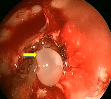
Clinical case: A 63-year-old male patient with racemose neurocysticercosis, hydrocephalus and cerebrospinal fluid fistula. He underwent transnasal endoscopy, removal of cysts from the sphenoid sinus, sellar, suprasellar, and prepontine regions, and fistula closure. He also presented erosion in the temporal bone and dural fistula, which were closed through microsurgery and endoscopy. The patient had a favorable initial evolution, with spastic quadriparesis, which improved with rehabilitation. Subsequently he presented episodes of ventriculoperitoneal shunt system dysfunction.
Conclusion: Neuroendoscopy is a diagnostic and therapeutic method of various forms of neurocysticercosis. Extraparenchymal neurocysticercosis is able to produce bone and dural erosion, so must be considered in the differential diagnosis of cerebrospinal fluid.
Keywords Neurocysticercosis, Fistula, Neuroendoscopy, Sphenoid sinus, Cysts (source: MeSH NLM)

