Usted está aquí
Peruvian Journal of Neurosurgery
Post-surgical subarachnoid hemorrhage in endonasal endoscopic resection of pituitary macroadenoma without arachnoid opening
GIAN FRANCO REYES N, OLENKA SAPALLANAY O, JERSON FLORES C, FERNANDO PALACIOS S.
Abstract (Spanish) ||
Full Text ||
PDF (Spanish)
ABSTRACT
Introduction: Pituitary adenomas represent 90% of tumors in the sellar region. The surgery is performed by transsphenoidal resection (TSR) or transcranial resection (TCR); TSR can be by microscopy or endonasal endoscopy. Subarachnoid hemorrhage (SAH) associated with a pituitary tumor is rare and can be: Preoperative, due to pituitary apoplexy with rupture of the arachnoid into the basal cisterns, Intraoperative, due to vascular injury during surgery or rupture of an unidentified aneurysm; and Postoperative, due to residual tumor hemorrhage, or bleeding from small vessels adhered to the tumor capsule.1, 2
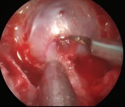
Clinical case: 46-year-old male patient with a clinical picture of bitemporal hemianopsia with left predominance. Magnetic resonance imaging and brain tomography showed a pituitary macroadenoma. Endoscopic endonasal resection of the tumor was performed without intraoperative opening of the arachnoid. On the 1st day, the evolution was favorable with improved visual fields, but on the 2nd day, the patient presented headache and increased visual deficit. Brain CT showed SAH in basal cisterns. Tomography angiography showed no vascular injury. The patient evolved favorably with the improvement of visual fields.
Conclusion: Subarachnoid hemorrhage associated with a pituitary adenoma has various causes that can be preoperative, intraoperative, and postoperative. The incidence of postoperative SAH as a complication of endonasal endoscopic resection is low, but it can affect the patient's prognosis. Excessive traction on the tumor capsule should be avoided.
Keywords: Subarachnoid Hemorrhage, Pituitary Neoplasms, Visual Fields, Arachnoid (source: MeSH NLM)