Usted está aquí
Peruvian Journal of Neurosurgery
High-definition three-dimensional exoscopy-guided exeresis of a ruptured supratentorial cavernous angioma. Initial experience at the Dos de Mayo National Hospital.
JOSÉ LUIS ACHA S., LUIS CONTRERAS, MIGUEL AZURÍN, MANUEL CUEVA, ADRIANA BELLIDO; SHAMIR CONTRERAS
Abstract (Spanish) ||
Full Text ||
PDF (Spanish)
ABSTRACT
Introduction: We report the exeresis of a cerebral cavernoma, by using the extracorporeal telescope ("exoscope") operating system integrated into the Kinevo 900 microscope. The objective of this study was to evaluate the surgical potential of this novel high-speed 3D exoscope system. definition (4K-HD) for the removal of a brain cavernoma.
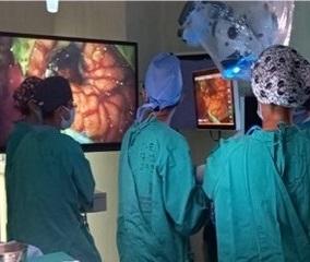
Clinical Case: A 47-year-old female patient was admitted to the emergency room for having presented intense headache with occipital predominance, the tomography showed a right occipital hematoma, the presence of a ruptured cerebral cavernoma was confirmed, so surgery was decided. The exoscope allowed good maneuverability of the instruments, without visual obstruction. The large 4K monitor made for an immersive surgical experience, giving multiple team members the same high-quality 3D view as the primary operator. It was also ergonomically favorable, allowing the surgeon to be in a neutral position regardless of the operative angle.
Conclusion: The novel system provided excellent visualization, ergonomics, and favorable maneuverability, the shared surgical vision of the exoscope with 3D lenses provided educational advantages for our residents. More cases are justified to validate this initial experience.
Keywords: Hemangioma, Cavernous, Central Nervous System, Telescopes, Ergonomics, Brain. (Source: MeSH NLM


