Usted está aquí
Peruvian Journal of Neurosurgery
Spontaneous intraventricular pneumocephalus associated with transient aphasia: case report and review of the literature
ZINDYA BARRRIENTOS M., CAMILO CONTRERAS C.
Abstract (Spanish) ||
Full Text ||
PDF (Spanish)
ABSTRACT
Introduction: Pneumocephalus or the presence of air in the cranial cavity is common after a craniotomy and in patients with traumatic brain injury; however, its spontaneous appearance is extremely rare. So far, very few cases of spontaneous intraventricular pneumocephalus have been reported. We present the case of a patient who developed spontaneous intraventricular pneumocephalus, which required a craniotomy for surgical correction.
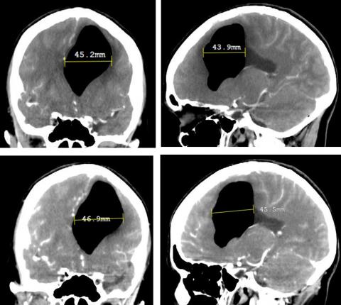
Clinical Case: A 59-year-old female patient with a history of left suboccipital craniotomy and resection of the left vestibular Schwannoma who after 2 years presented headache of 3 weeks duration and aphasia of expression. On examination: Mild expressive aphasia, surgical scar with no evidence of cerebrospinal fluid (CSF) leakage. Brain tomography (CT) showed pneumoventricle in the frontal and temporal horns of the left lateral ventricle and deviation from the midline; radionuclide cisternography was negative for CSF fistula and CSF analysis was normal. A left subtemporal craniotomy was performed, finding a bone defect in the petrous portion of the temporal bone above the internal auditory canal associated with a meningocele of the medial base of the skull, which was sealed with bone wax, fat, fascia lata, and biological glue.
Conclusion: The first case of spontaneous intraventricular pneumocephalus without identifiable CSF fistula is described, which made this case extremely rare. The treatment performed was a surgical correction of the meningocele through a subtemporal extradural approach, and the patient presented a favorable evolution with the improvement of the aphasia.
Keywords: Pneumocephalus, Aphasia, Meningocele, Craniotomy, Temporal Bone (source: MeSH NLM)