Usted está aquí

Jan-Mar 2022, Volume 4, Issue 1

Prognostic factors in the survival of patients operated of astrocytoma grade III at the Guillermo Almenara Hospital Lima- Peru. 2003-2009

Enviado por admin el Lun, 06/20/2022 - 02:15
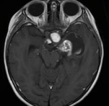
Anaplastic astrocytoma (AA) or grade III is a primary brain tumor, astrocytic, malignant, and diffusely infiltrating. The survival of patients depends on several clinical and treatment factors, being this unknown in our environment. The objective of this study was to determine the survival of patients operated on for grade III astrocytoma and the impact of preoperative and postoperative prognostic factors.

Glioma of the optic pathway and hypothalamus in a child: a case report

Enviado por admin el Lun, 06/20/2022 - 02:03

Gliomas of the optic nerve, visual pathway, and hypothalamus are treated as a single entity, being considered benign neoplasms in pediatric age, grade I according to the WHO. 25% of them are confined to the optic nerve, 40-75% involve the optic chiasm, and 33-60% are posterior lesions. Most do not present symptoms, but in the case of presenting, the most frequent is loss of vision. The gold standard for diagnosis is magnetic resonance imaging (MRI) with contrast. The first line of treatment is chemotherapy, with surgery used for nerve decompression, if necessary.

Esthesioneuroblastoma with intracranial invasion, surgical management by double approach: craniotomy and craniofacial. case report

Enviado por admin el Lun, 06/20/2022 - 01:57

Esthesioneuroblastoma, or olfactory neuroblastoma, is a rare malignant neoplasm of the sinonasal tract that originates in the olfactory neuroepithelium with neuroblastic differentiation. It occurs most frequently in the upper nasal cavity. It is a locally aggressive neoplasm and metastasizes both hematogenously and lymphatic. A multimodal approach, which can combine surgery, chemotherapy, and radiotherapy, is essential for the management of these tumors.

Post-surgical subarachnoid hemorrhage in endonasal endoscopic resection of pituitary macroadenoma without arachnoid opening

Enviado por admin el Lun, 06/20/2022 - 01:47

Los adenomas de hipófisis representan el 90% de los tumores de la región selar. El cirugía se realiza mediante la resección transesfenoidal (RTE) o transcraneal (RTC); La RTE puede ser mediante microscopía o endoscopía endonasal. La hemorragia subaracnoidea (HSA) asociada a un tumor de hipófisis es rara y puede ser: Preoperatoria, por apoplejía hipofisiaria con ruptura de aracnoides hacia las cisternas basales, Intraoperatoria, debido a una lesión vascular durante la cirugía o ruptura de un aneurisma no identificado; y Postoperatoria, debido a hemorragia del tumor residual, o sangrado de pequeños vasos adheridos a la cápsula tumoral.

Spontaneous intraventricular pneumocephalus associated with transient aphasia: case report and review of the literature

Enviado por admin el Lun, 06/20/2022 - 01:41

Pneumocephalus or the presence of air in the cranial cavity is common after a craniotomy and in patients with traumatic brain injury; however, its spontaneous appearance is extremely rare. So far, very few cases of spontaneous intraventricular pneumocephalus have been reported. We present the case of a patient who developed spontaneous intraventricular pneumocephalus, which required a craniotomy for surgical correction.

High-definition three-dimensional exoscopy-guided exeresis of a ruptured supratentorial cavernous angioma. Initial experience at the Dos de Mayo National Hospital.

Enviado por admin el Lun, 06/20/2022 - 01:32

We report the exeresis of a cerebral cavernoma, by using the extracorporeal telescope ("exoscope") operating system integrated into the Kinevo 900 microscope. The objective of this study was to evaluate the surgical potential of this novel high-speed 3D exoscope system. definition (4K-HD) for the removal of a brain cavernoma.

Giant occipital encephalocele in a newborn; surgical treatment. Case report

Enviado por admin el Lun, 06/20/2022 - 01:21

Neural tube defects (NTD) are congenital malformations that are caused by the lack of fusion of the neural tube during the embryonic period, exposing the nervous tissue to the outside. There are different types, and they can be cranial (anencephaly and encephalocele) and spinal (spina bifida). The encephalocele is a protrusion or herniation of the intracranial content, through the bony defect of the skull. In this article, we report the case of a patient diagnosed with occipital encephalocele, while reviewing the neurosurgical treatment performed in our hospital.

Suscribirse a RSS - Jan-Mar 2022, Volume 4, Issue 1