Usted está aquí
Peruvian Journal of Neurosurgery
Magnetic Resonance Tractography integrated to Neuronavigation in the surgical planning of a temporal arteriovenous malformation at the Dos de Mayo National Hospital. Case report.
JOSÉ LUIS ACHA S., MIGUEL AZURÍN, ADRIANA BELLIDO.
Abstract (Spanish) ||
Full Text ||
PDF (Spanish) ||
PDF (English)
ABSTRACT
|
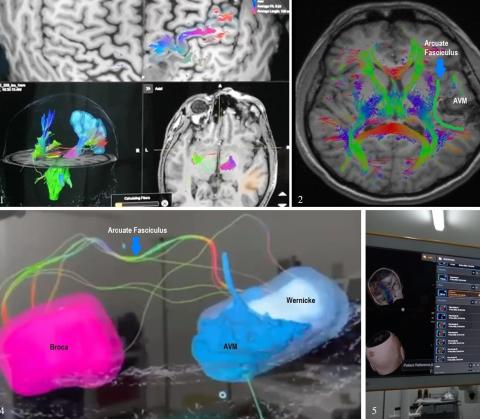
Introduction: Complete resection of a cerebral arteriovenous malformation (AVM) eliminates the risk of bleeding1. Although AVMs that adjoin eloquent areas have been studied with functional neuroimaging or intraoperative mapping, 2 the usefulness of tractography has been limited to case reports or small series. Selecting the patient for surgery for an AVM close to an eloquent area is a challenge. 3 Clinical case: 33-year-old man with a clinical picture of epilepsy for 8 years controlled with carbamazepine. Two years ago, after suspension of treatment, the seizures reappeared, some of the auditory hallucinations "voices asking for help." Brain tomography (CT) showed a hyperdense lesion suggestive of AVM in the left temporal region, which was confirmed with magnetic resonance imaging (MRI) and cerebral angiography. The AVM was completely resected using the tractography integrated into the Neuronavigation. Conclusion: Magnetic resonance tractography integrated into the Neuronavigation allows to assess in real-time the proximity of the nidus of AVM to the arcuate fasciculus tract and the use of intraoperative fluorescein video angiography allows to assess vascularity in real-time. All of this makes it possible to perform total resection without causing injury to the eloquent area by avoiding compromising the fibers of the arcuate fasciculus tract. Keywords: Intracranial Arteriovenous Malformations, Neuronavigation, Fluoresceins (Source: MeSH NLM) |