Usted está aquí
Peruvian Journal of Neurosurgery
Successful management of ruptured cavernous malformation of the pons
JOHN VARGAS U., FERNANDO PALACIOS S., EDUARDO ROMERO V.
Abstract (Spanish) ||
Full Text ||
PDF (Spanish) ||
PDF (English)
ABSTRACT
Introduction: Brain stem cavernomas constitute 18-35% of intracranial cavernomas and have the highest bleeding rate of all brain cavernomas. Its annual rebleeding rate is 21 to 60%. Asymptomatic injuries should be treated conservatively, while symptomatic and accessible injuries surgery is recommended. Surgical resection prevents neurological deterioration caused by recurrent bleeding.
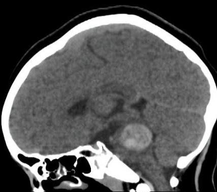
Clinical Case: We present the case of a 10-year-old female patient with headache, ataxic gait, left LV PC paresis, and vertigo. A magnetic resonance imaging of the brain (MRI) was carried out, where a ragged expansive process was evidenced in T2 in the posterior protuberance region with intra-tumoral and extra-tumoral hemosiderin deposits. Brain angiography was negative. A medial suboccipital craniotomy with a telovelar approach was performed, achieving total cavernoma resection. The patient was extubated on the 2nd postoperative day. Non-contrast brain tomography (CT) showed no acute complications, only minimal bleeding from the operative bed. Post-surgical brain MRI showed the absence of the lesion. At 7 months, she continued without motor deficit, with adequate gait and Glasgow 15 points.
Conclusion: Brain stem cavernomas are a rare disease with high morbidity. Proper patient choice, surgical approach, and time to surgery decrease post-surgical morbidity.
Keywords: Hemangioma, Cavernous, Central Nervous System, Brain Stem, Craniotomy (Source: MeSH NLM)