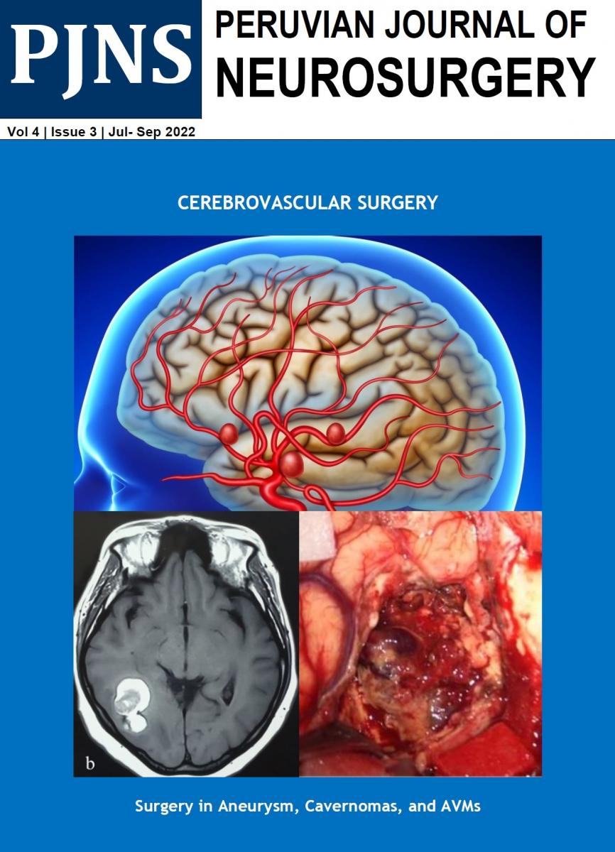
Objective: To determine the validity, sensitivity and specificity of multislice spiral CT angiography (ATEM) with a 16-Multislice CT scanner detector lines in the diagnosis of cerebral aneurysms and their most frequent localizations.
Patients and Methods: We selected 51 patients with nontraumatic subarachnoid hemorrhage due to suspected intracranial aneurysm who underwent surgery in the Hospital Daniel A. Carrión, Callao, for the period May 2004 to November 2007.
Results: The sensitivity of the ATEM for the detection of intracranial aneurysms in this study was 100%. The resulting specificity was 50%. The positive predictive value (PPV) of ATEM for the diagnosis of cerebral aneurysms was 98%, with 87.8% accuracy of the method for determining the location of the aneurysm and 12.2% of uncertainty in the determination of the location of this anomaly. The average size of the aneurysm at surgery was 11.33 + / - 6.9 mm. While the average size of the aneurysm by ATEM was 8.60 + / - 5.75 mm. The difference between the means of these measurements was significant (p = 0.027). 70.3% of aneurysms in this study were found in posterior communicating segment of both internal carotid and middle cerebral arteries both.
Conclusion: CT angiography is an excellent noninvasive test for the detection of intracranial aneurysms and to determine its location.
Key words: Intracranial aneurysm, CT Angio (ATEM), aneurysm surgery
Objetivo: Determinar la validez, así como la sensibilidad y especificidad de la angiografía por tomografía espiral multicorte (ATEM) con un tomógrafo Multicorte de 16 líneas de detectores, en el diagnóstico de los aneurismas cerebrales y las localizaciones más frecuentes.
Pacientes y Métodos: Se seleccionaron 51 pacientes que tenían hemorragia subaracnoidea no traumática y que se sospechaba de aneurisma intracraneal, operados en el Hospital Daniel A. Carrión del Callao en el periodo de mayo del 2004 a noviembre del 2007.
Resultados: La sensibilidad de la ATEM para la detección de aneurismas cerebrales, en el presente estudio fue de 100%. La especificidad resultante fue de 50%. El valor predictivo positivo (VPP), del ATEM para el diagnóstico de los aneurismas cerebrales, fue de 98%, con una certeza de 87.8% del método para determinar la localización del aneurisma, y un 12.2% de imprecisión en la determinación de la localización de esta anomalía. El tamaño promedio del aneurisma en la cirugía fue de 11.33 +/- 6.09 mm. Mientras que el tamaño promedio del aneurisma por ATEM fue de 8.60 +/- 5.75 mm. La diferencia entre las medias de estas mediciones fue significativa (p=0.027). Un 70.3% de los aneurismas en este estudio se hallaron en segmento comunicante posterior de ambas carótidas internas y ambas arterías cerebrales medias.
Conclusion: La angiotomografía es una excelente prueba no invasiva para la detección de aneurismas de intracraneales y determinar su ubicación.


