Usted está aquí
Peruvian Journal of Neurosurgery
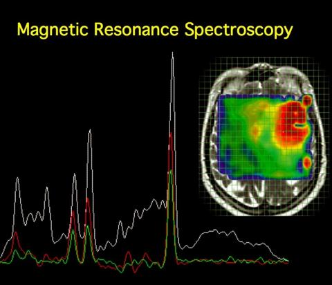
MRI Spectroscopy (H+) in the differential diagnosis between brain tuberculomas and intra-axial neoplastic process
Carlos Martinot L. MD, Carlos Martinot Del P. MD, Genaro Herrera G. MD, David Alfaro L. MD, Augusto Merello K. MD, et Al.
Abstract (Spanish) ||
Full Text ||
PDF (Spanish)
ABSTRACT
Objective: To study changes in brain metabolites between brain tuberculomas and intra-axial neoplastic process so that we allow an approach to its diagnostic differentiation.
Patients and Methods: A descriptive case series, 24 patients aged between 02 and 72 years who underwent the brain structural and spectroscopic study univoxel with voxel of 20 mm x 20 mm x 20 mm (8 cc ) located at the level of the lesion determined. Resonator was used 1.5-tesla General Electric Smart Speed and Hdx models. Ratios were assessed Choline / creatine, and N-Acetil-Aspartato/Creatina and Mio-inositol/Creatina. All cases were confirmed anatomical - pathological or therapeutic test.
Results: The ratio choline / creatine for intra-axial brain tumors was found to be 1.97 compared with tuberculomas which was 1.15, which is statistically significant (p = 0.017, Mann Whitney). Mio-inositol/creatina ratio for intra-axial brain tumors was 0.89 compared with tuberculomas was 0.55 which is statistically significant (p = 0.008, Mann Whitney). The ratio N-Acetil-Aspartato/Creatina for intra-axial brain tumors was 0.96 compared with tuberculomas was of 1.28 which is statistically significant (p = 0.003, Mann Whitney).
Conclusion: Magnetic resonance spectroscopy allows a more accurate diagnosis of intraparenchymal brain lesions of uncertain etiology and occurs when the lesion is unique, brain neoplasia and tuberculomas, enabling better-targeted treatment for patients.
Key words: Nuclear Magnetic resonance, MR espectroscopy, tuberculoma, brain tumors.